eGFP mRNA: A Revolutionary Tool for Tracking Gene Expression
The Cy5 labeled mRNA and eGFP expression patterns observed with different delivery techniques reveal essential information about how mRNAs are trafficked in the skin. For example, after intradermal injection, eGFP expression was seen in dermal fibroblasts, epithelial cells of appendageal structures (porous skin units and sweat glands), and vascular tissue. Similarly, after epidermal fractional laser ablation, eGFP expression was observed in keratinocytes near the pores and skin surface but not deeper within the dermis or into vascular or lymphatic tissues.
Background
GFP (green fluorescent protein) is a marker gene for tracking cell movements and local protein expression. It is derived from the jellyfish Aequorea victoria and binds to a protein called aequorin, which emits blue luminescence in the presence of Ca2+ ions. GFP fluorescence is induced by excitation of the protein, so it can be visualized in living cells without exposing them to toxic chemicals. A commercial capped eGFP mRNA product mimics fully processed mature mRNA. This stable mRNA has a natural 509-nm emission peak, making it an excellent tool for tracking protein expression. This approach showed that a significant fraction of 34-DCs express eGFP at 3 hours following mRNA electroporation. In addition, smFISH analysis confirmed that eGFP is expressed explicitly in these cells. The expression kinetics are comparable to those seen in K562 cells following transgene expression by a viral vector.
Moreover, the absence of co-localization between the mRNA signal and eGFP fluorescence near the injection site supports the hypothesis that eGFP is locally expressed by keratinocytes and epithelial cells of skin appendages. However, the mechanism by which mRNA reached these cells remains unknown. It could be through reflux triggered by high back-pressure emitted during the injection process or passive migration from the deeper dermis to the sample periphery via lymphatic and blood vessel networks in this layer.
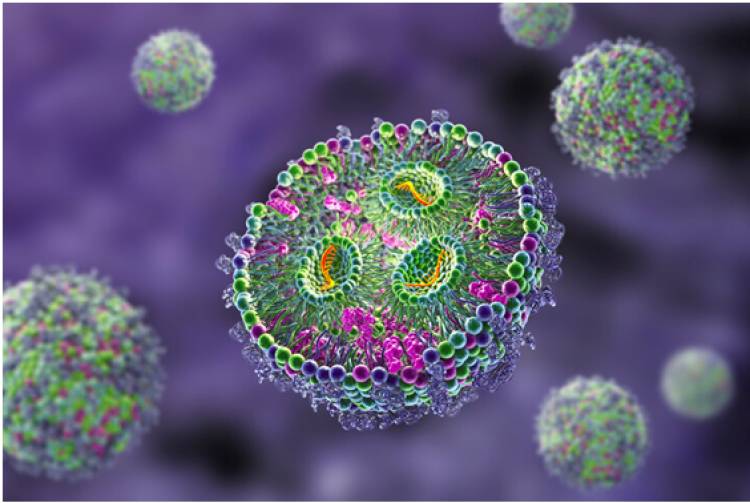
Methods
A simple reporter gene system was used to directly express a fluorescent protein (GFP) in the cytoplasm to visualize cell protein expression. mRNA encoding eGFP is easy to prepare and can be quickly delivered into cells using standard nucleofection methods. The eGFP protein is exceptionally bright and can be easily tracked by fluorescence microscopy. It also has a characteristic excitation maximum of 488 nm and an emission maximum of 509 nm, which makes it ideal for tracking protein expression. First, nucleofection was verified to successfully deliver two nL lipoplexes containing free mCherry mRNA, eGFP mRNA, or a 50/50 mixture of both into mammalian cells. We found that mRNA delivery and protein expression were highly correlated and could be detected quickly after injection. Using the same method, 34-LCs were electroplated with plasmid DNA constructs encoding eGFP, Melan-A, or luciferase. It was observed that mRNA distribution and eGFP protein expression co-localized mainly in the epidermis but not in the dermis of skin samples. A single-chain fused-to-eGFP (smFISH) probe that recognizes eGFP transcript explicitly and can be labeled with Cy5 was generated for more detailed analysis. smFISH reveals both eGFP protein and eGFP mRNA in the same cell. We observed that eGFP mRNA expression was more prevalent in the epidermis than the dermis and correlated with keratinocytes.
Results
In addition to being used as a quantitative reporter gene, GFP also reveals molecular pathways that regulate protein expression. In the present study, eGFP mRNA testing was performed using HEK-293T cells (ATCC CRL-3216) transfected with mRNA-LNP complexes and analyzed by widefield and confocal fluorescent microscopy. The test results showed that mRNA-LNPs could effectively transfect HepG2 spheroids and induce eGFP expression. The data also indicate that LNP(DSPC) and LNP(DOPE) transfect HepG2 spheroids with similar potency. However, eGFP expression is reduced by LNP(DOPE) transfections, which may be due to mRNA internalization. As shown by high-content imaging, Cy5-tagged mRNAs are rapidly internalized and quickly escape from the LNP and endosome. However, mRNA that remains trapped inside the LNP or endosome would not be translated into functional proteins. Flow cytometric analyses of eGFP expression after mRNA-LNP transfections of Mo-DCs revealed that the various delivery techniques affect cell type distribution and site of eGFP expression. EGFP expression is primarily driven by dermal fibroblasts and, to a lesser extent, epidermal keratinocytes in appendageal structures, sweat glands, and vascular cells.
Moreover, using eGFP mRNA to analyze cellular uptake and trafficking of an experimental therapeutic provides a valuable tool for designing targeted mRNA-based drugs. The results presented here show that a combination of an automated workflow, a flow cytometer capable of measuring GFP fluorescence, and a customizable mRNA-LNP library offers a highly flexible approach to mRNA testing.
Conclusions
Compared to pDNA, mRNA-mediated transfection exhibits distinct single-cell expression kinetics. This enables a simple but accurate calibration of FC detection. Using microchannels with known dimensions that are filled with eGFP solutions of different concentrations, a time-lapse movie is recorded, and the grey values of the images are analyzed to produce a calibration curve. Using this, the number of successfully delivered and translated mRNA molecules can be calculated accurately. Consequently, the dose-response relation of mRNA-mediated gene regulation can be reproduced.
Moreover, the simultaneous measurement of Cy5-tagged mRNA internalization and eGFP translation by high-content imaging reveals that these processes are highly linear. Especially for mRNA doses close to the EC50 value, the total green object area increases rapidly up to 7.5 h post-transfection. In contrast, the onset of eGFP expression with pDNA is spread over several hours. This new tool combines the advantages of a flow cytometer with a powerful image analysis software package to provide an easy-to-use system for high-throughput GFP fluorescence measurements. To ensure reliable results, all workflow files are tested with a "test" file, and the tubes are rinsed twice with RNase ZAP and three times with DEPC-treated water. In addition, the mRNA is prepared according to a specific protocol that automatically adjusts reagent volumes and flow cytometer settings. The resulting data are easily visualized and exported in standard formats.